S-Sharp
>
Applications
>
Preclinical Imaging
>
Developmental Biology
Preclinical Imaging
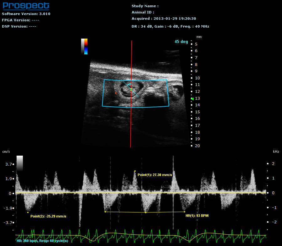
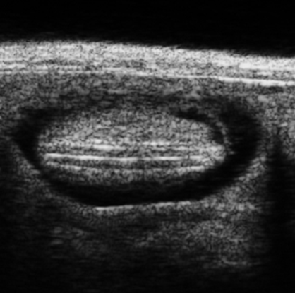
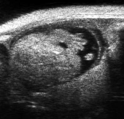
With complete modes including color Doppler, Prospect allows visualization of rodent embryonic development even in the early pregnancy stage. Structures such as embryo cells, chambers, mitral valve, and vessel flows can be identified and quantified.
| E10.5 mouse embryos | E10.5 mouse embryo |
| Embryonic cardiology in DP mod | E14.5 embryo (M mode) |
| E7.5 mouse embryos | E12.5 embryonic brain ventricle |
 |
||
| E9.5 mouse embryonic heart and neural tube | E9.5 mouse embryo (PW mode) |
 |
 |
|
| E12.5 mouse embryonic spinal cord | E14.5 mouse embryonic head and forelimb |
| E13.5 Embryonic umbilical cord in DP mode |
|
| E14.5 embryo image in coronal plane |
E14.5 embryo image in sagittal plane |
| E14.5 embryo image in transverse plane |
E14.5 embryo (PW mode) |
| E14.5 embryonic lateral ventricle injection |
E15.5 embryonic lateral ventricle injection |