|
|
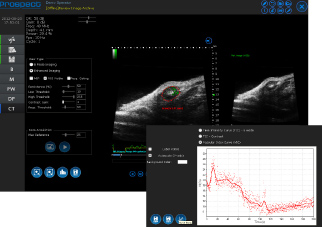
Doppler mode |
|---|---|
|
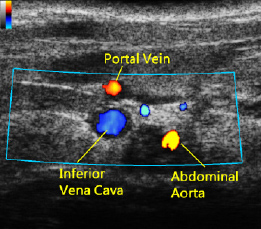
Three kinds of Doppler modes are provided: color Doppler mode, power Doppler mode, and Pulsed-wave Doppler mode. |
|
|
Ultrasound cross-sectional imaging of the mouse portal vein, abdominal aorta, and inferior vena cava in color Doppler mode. |
B mode |
 |
|---|---|
|
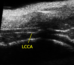
2D gray-level imaging is displayed in real time with the improved lateral resolution using the Virtual Array technology. ECG-triggered, respiration gated imaging is achieved for ultrahigh frame rates. | |
|
Longitudinal axis view of the left common carotid artery (LCCA) in B mode. |
3D Imaging mode |
|---|
|
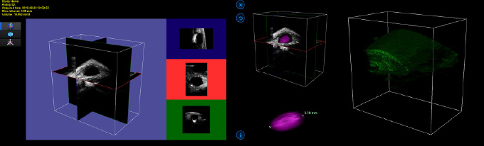
3D image visualization, reconstruction, and volumetric measurements are provided. |
 |
|
3D imaging of breast cancer for volumetric measurements. |
M mode |
 |
|---|---|
|
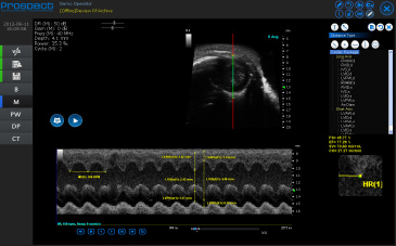
M mode assesses motion information as a function of time. Dimension changes of the target organ can be analyzed. | |
|
Wall thickness and cardiac function measurements of the left ventricle (short axis view) in M mode. |
|
|
Contrast mode |
|---|---|
|
Microbubble based contrast imaging allows measurements of time-intensity curve(TIC), vascular index curve (VIC) and data export for off-line analysis. |
|
|
Perfusion image of a mouse tumor in the contrast mode.Vascular index curve is also displayed. |
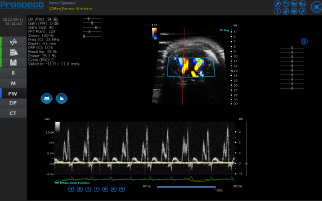
Pulsed-wave (PW) Doppler mode |
 |
|---|---|
|
PW Doppler shows blood flow velocity profile as a function of time. The duplex or triplex mode allows the users to identify flow locations and simultaneously conduct quantitative flow measurements. | |
|
Doppler spectrum of blood flows at the mouse tricuspid in the four chamber view is shown. |
|
|
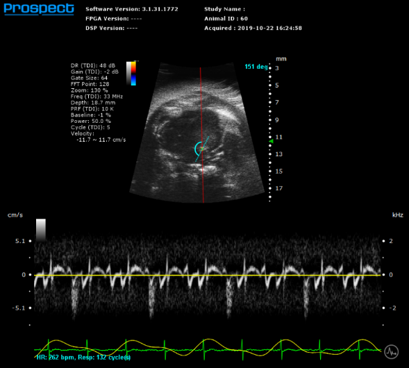
Tissue Doppler mode |
|---|---|
|
Tissue Doppler imaging is used to measure the low-velocity, high-intensity signals of the myocardium. |
|
|
Tissue Doppler imaging at mitral annulus. |
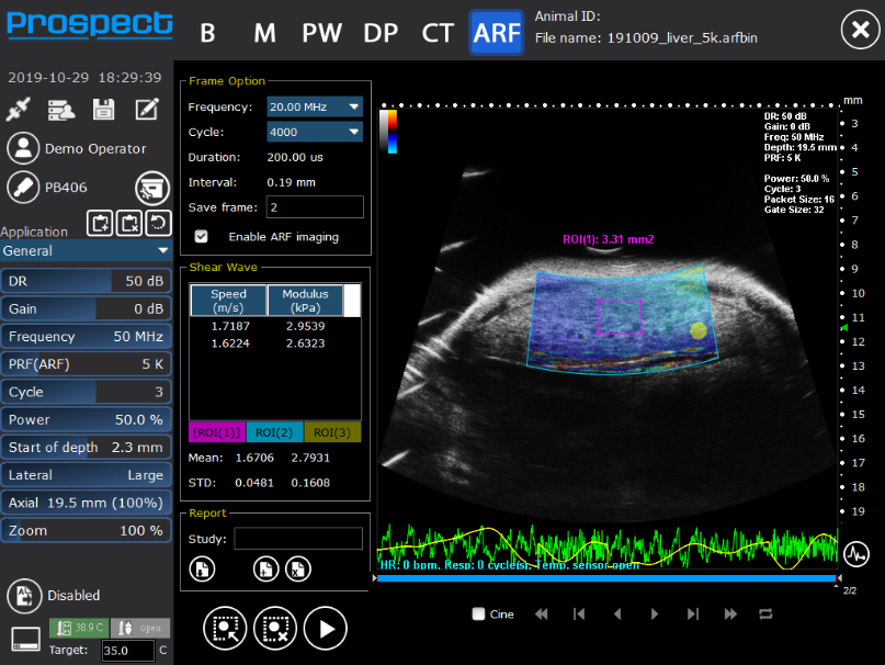
ARF mode (Elasticity Imaging) |
 |
|---|---|
|
Shear wave elasticity imaging is a non-invasive method of measuring the elasticity of tissues. With the confocal design, shear wave propagation and velocity can be detected and calculated. | |
|
Shear wave elasticity imaging of liver. |
|